
The chest x-ray view shows a widened mediastinum.
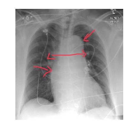
The above chest x-ray, in the clinical context is concerning for a possible aortic dissection. The x-ray shows widened mediastinum, double aortic contour & an outward bulge in the area of ascending aorta.
The patient had Stanford A aortic dissection on CT aortogram and underwent immediate surgery.
Chest x-ray features are nonspecific in a patient with aortic dissection. Following abnormalities could be present.
- Mediastinal widening.
- Double aortic knob sign.
- Enlarged aorta with poorly defined aortic contour.
- Inward displacement of the aortic wall calcification by more than 10mm (calcium sign).
- Cardiac enlargement (pericardial effusion).
- Left sided pleural effusion.
- Left apical opacity (apical cap).
- Tracheal displacement to the right.
- Depression of the left main bronchus.
Further reading https://emedicine.medscape.com/article/416776-overview#a2