
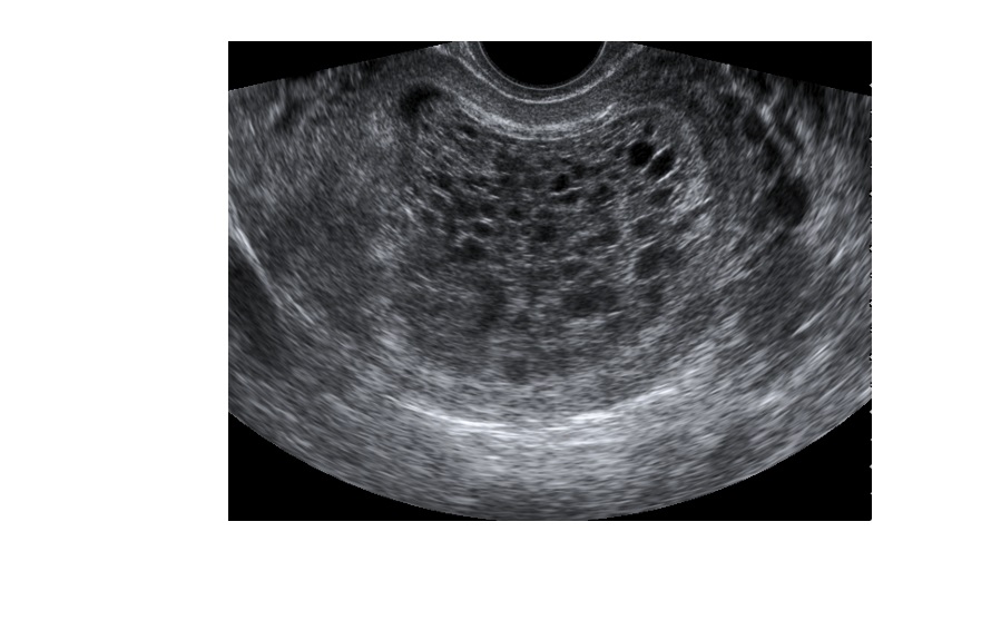
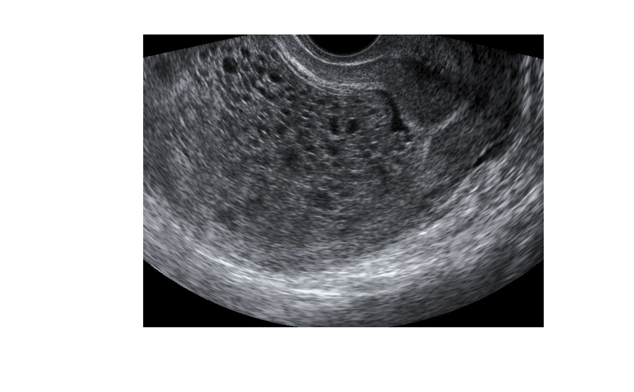
The following ultrasound images are from a 38 year old, with a h/o 8 weeks LMP, who presented with PV bleed. What radiological sign can you see in the images?
click to enlarge
click to enlarge
The ultrasound images (transverse and longitudinal) show multiple cysts occupying the uterine cavity, giving rise to the ‘bunch of grapes sign’. This finding is suggestive of a molar pregnancy. There is no intrauterine gestational sac seen.

click to enlarge
The patient had a very high serum quantitative hCG. She did not have any evidence of metastasis upon further tests. She underwent dilatation and curettage.
Molar pregnancy:
- More common at the extremes of reproductive age.
- Complete mole (no fetal tissue) or partial mole (fetal tissue present).
- Clinical presentation: PV bleeding, hyperemesis, hyperthyroidism (due to stimulation of the thyroid by high circulating hCG or production of thyroid stimulating substance by the trophoblastic tissue), pre-eclampsia.
- Very high quantitative hCG usually, but a normal hCG level is also a possibility.
- Baseline chest radiography must to r/o metastasis.
Reference – http://emedicine.medscape.com/article/254657
[/peekaboo_content]