
The following OPG x-ray is from a 37 year old male who has presented with a fever as well as pain and swelling over his left neck. He has experienced left sided neck pain related to meals for the last few months. What can you see on the x-ray?
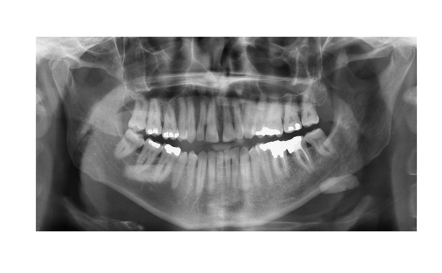
click to enlarge
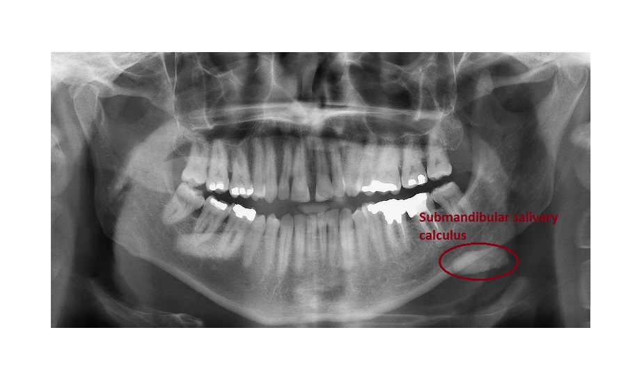
The OPG view shows a large calculus (25x10mm) over the left submandibular area, which is suggestive of a submandibular stone.
click to enlarge
Ultrasound of the submandibular area confirmed a large stone in the left submandibular duct with associated obstruction and inflammation in the gland.

click to enlarge
This patient had developed acute sialadenitis due to an ascending infection from the blocked duct.
He was commenced on intravenous antibiotics and was referred to ENT services for further management (stone extraction).
About 70% of submandibular calculi are radio-opaque. Calculi are more common in the submandibular gland compared to the parotid due to non-dependent drainage. Also, increased viscosity of the secretions in the SM gland accounts for a higher incidence of stones.
Reference: http://emedicine.medscape.com/article/882358
[/peekaboo_content]