
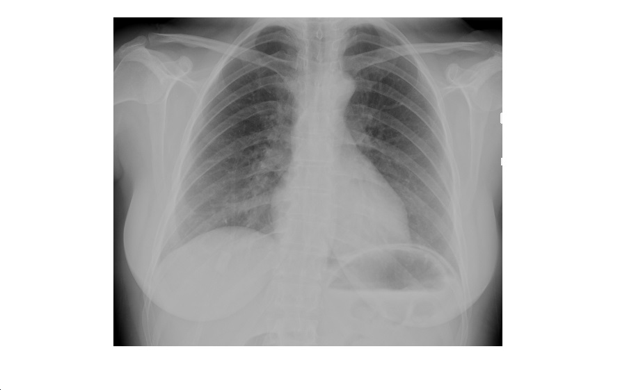
This PA erect chest x-ray is from a 40 year old female patient who has presented with abdominal pain. What can you deduce from the x-ray?
click to enlarge
The lung fields are clear and the cardiomediastinal contour is normal. However, there appears to be an air crescent under the left diaphragm. Is this pneumoperitoneum?
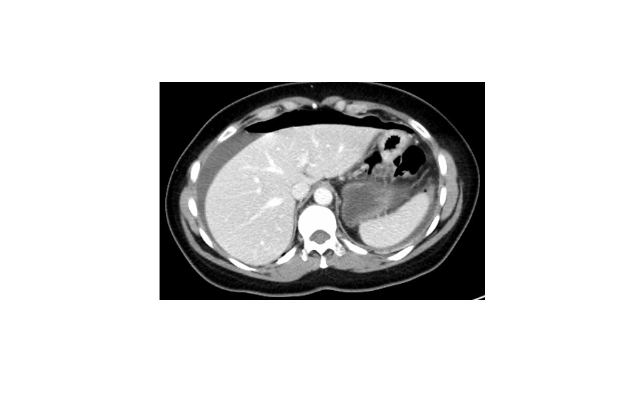
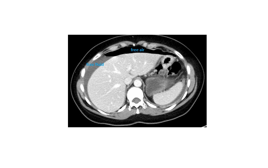
The patient indeed had a large pneumoperitoneum, secondary to a perforated antral ulcer; the following CT scans were obtained just an hour after the plain x-ray!
click to enlarge
click to enlarge
Pneumoperitoneum:
- About 76% of perforated peptic ulcers will reveal free air on erect chest x-rays.
- In patients who are unfit or too sick to stand upright, a left lateral decubitus x-ray can show a small pneumoperitoneum.
- Both erect cxr and left lateral decubitus abdominal x-rays require the patient to be in position for at least 10 mins to allow sufficient time for the air to rise.
- On the left, it can be difficult to distinguish free air from gas in the stomach/colon.
- CT is the most sensitive investigation for the detection of free peritoneal gas.
(Reference: Textbook of Radiology and Imaging by Sutton)
[/peekaboo_content]