This ECG is typical posterior MI with deep horizontal STD in V1-V3. Patient was transferred to SCGH cathlab priority 1, loaded with Aspirin 300 mg and Heparin 5000 U iv as per their cardiology advice.
Posterior STEMI:
- Isolated 3-11 %
- Usually in context of an inferior or lateral STEMI
- Horizontal STD
- Tall, broad RW > 30 ms
- Upright TW
- R/S >1 in V2
The progressive development of pathological R waves in posterior MI mirrors the development of Q waves in anteroseptal STEMI
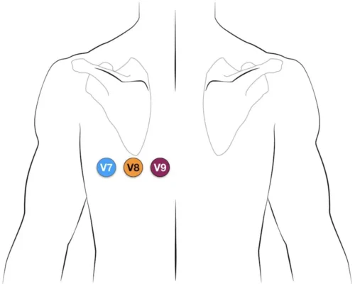
Perform V7-V8-V9 ECG – shows STE – only 0.5 mm is enough to diagnose STEMI.
See the attached file for posterior leads placement.

Thanks to Dr Svetlana Trandos for preparing this case
