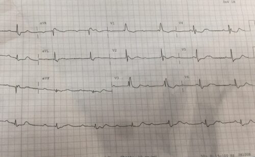
A 64 year old male presents to ED after multiple seizure episodes that day. It was reported that the patient would have 20 seconds of seizure activity and then make a full recovering. The patient has a history of IHD. At time of ECG the patient was symptom free:
Describe and interpret the ECG
Answer:
Rate: Ventricular rate 42 beats/min Atrial rate 90 beats/min
Rhythm: AV dissociation – third degree heart block
Axis: Left axis
Intervals:
PR –
QRS 120ms
QT 469ms (difficult to accurately assess due to P waves within QRS and T waves
Additional:
No Peaked T waves, ST depression V4-6
The above ECG shows a complete heart block with a slow ventricular escape rhythm. There is some ST depression suggestive that ischemia is the likely cause, however drugs and electrolyte abnormalities especially hyperkalaemia needs to be excluded.
The patients “seizure” episodes correlated to episodes of ventricular standstill on cardiac monitoring. The patient was externally paced and started on an isoprenaline infusion and then had a PPM inserted

