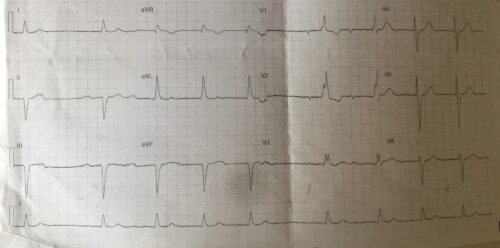
A 83 year old male presents to ED after a syncopal episode. Below is the patients ECG
Describe and Interpret the ECG
Answer
Rate: 54 beats per minute
Rhythm: Mobitz Type I Second Degree AV block
Axis: Left Axis
Intervals:
PR: Increasing PR segment, followed by drop QRS
QRS: 160ms
QTc: 455ms
Additional:
RBBB Morphology
Left Anterior Fascicular Block noted by left axis, qR wave I and aVL, rS wave in II, III, aVF
The combination of RBBB, LAFB and second degree block indicates a possible incomplete trifascicular block. As second degree AV blocks can occur at the AV node and therefore might not involve fascicular disease, so a diagnosis of a trifascicular block can only be made through a His Bundle recording.
In the context of syncope a Mobitz Type I Second degree AV block alone as well as a possible trifascicular block requires referral and admission under cardiology.

