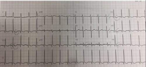
A 24 year old Indigenous female P2 G3 31/40 pregnant presents to ED with left sided non pleuritic chest pain and shortness of breath. Below is her ECG:
Describe and Interpret her ECG
Answer:
Rate: 108 beats per minute
Rhythm: normal sinus rhythm
Axis: Normal Axis
Intervals:
PR: 120ms
QRS: 90ms
QTc: 341ms
Additional:
- Inferolateral symmetrical T wave inversion
- Q waves Inferolaterally
- QRS complexes appear tall and deep, but strictly do not meet definition for LVH
The above ECG shows a sinus tachycardia with inferolateral T wave changes and Q waves. The Tall complexes do not meet criteria for LVH and the T wave inversion is symmetrical, and therefore less likely to be nonvoltage changes for LVH which usually show asymmetrical T wave inversion. LVH diagnosis needs to be excluded on echo
ECG changes in pregnancy are usually minor with sinus tachy and minor right or left axis shift. However some small studies have shown left axis deviation, Q waves inferiorly and T wave inversion in lead III,V1-V3.
In this clinical context the following pathology needs to be excluded as it can account for the ECG changes – PE, perimyocarditis, pregnancy induced cardiomyopathy, underlying structural disease from previous Rheumatic fever, ACS or spontaneous coronary artery dissection (SCAD)
The patient was admitted and had a normal echo, normal CTPA and normal serial troponins. The patient has further cardiology follow up as an out patient.
References
Sunitha, M. Electrocardiographic QRS axis, Q wave and T wave changes in 2nd and 3rd Trimester of normal pregnancy. Journal of Clinical and Diagnostic Research 2014 Sep 20
Sanghavi, M Cardiovascular Physiology of Pregnancy. American Heart Association Journal 2014

