
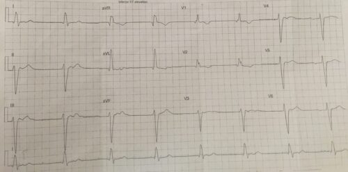
A 70 year old female presents to ED complaining of shortness of breath and chest pain. Below is the patients ECG:
Describe and Interpret the ECG:
Rate: Ventricular rate 48 beats per minute, Atrial rate complexes 1-3 and 5-7 48 beats per minute
Rhythm: Complexes 1-3 and 5-7, P waves occur in ST segment, complex 4 no associated P wave, complex 8 P wave before QRS ? associated to eachother
Axis: Left Axis
Intervals
- PR –
- QRS 200ms RBBB morphology
- QT 465ms (Bazett)
Additional:
Left Anterior Fascicular Block
1-1.5mm ST elevation inferolaterally
The above ECG shows a RBBB and LAFB with ST segment elevation in the inferolateral leads. With respect to the rhythm there appears that there might not be an association between the P waves and QRS complexes despite their similar rates. Complex 4 appears to be a escape rhythm with no P waves, with the same morphology as the other complexes, suggestive that complexes 1-3 and 5-7 could possibly be isorhythmic AV dissociation with a ventricular escape rhythm or a junctional escape rhythm with a RBBB. A longer rhythm strip would be required to further interpret the rhythm.
The ECG and clinical context is concerning for acute coronary syndrome. Other causes for this ECG would be electrolyte abnormalities and medication.
Chan TC, Brady WJ, Harrigam RA, Ornato JP, Rosen P, 2005, ECG in Emergency Medicine and Acute Care, Elsevier Mosby, USA
Amal Mattu’s ECGweekly.com