
The following lumbar spine AP x-ray is from a 55 year old woman with low back pain. What can be seen?
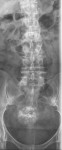
The single AP view looks normal; however, there is an incidental finding of popcorn calcification in the pelvis. Given its location in the pelvis, this is likely a calcified uterine fibroid.
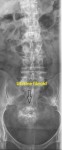
Uterine fibroid (also called leiomyoma):
- Benign neoplasm arising from the smooth muscle.
- Location within the uterus can be submucous, subserous or intramural
- They can be the cause of menorrhagia and lower abdominal pain
- Submucous and subserous myomas can be pedunculated and as such, can undergo torsion.
- Risk of malignant transformation is between 0.1-0.5% (leiomyosarcoma)
- Imaging modality of choice is ultrasound
- If symptomatic, will need appropriate outpatient referral