
These chest x-ray images are from a 39 year old heavy smoker who has presented with right-sided pleuritic chest pain and a fever. What can you glean from these images?
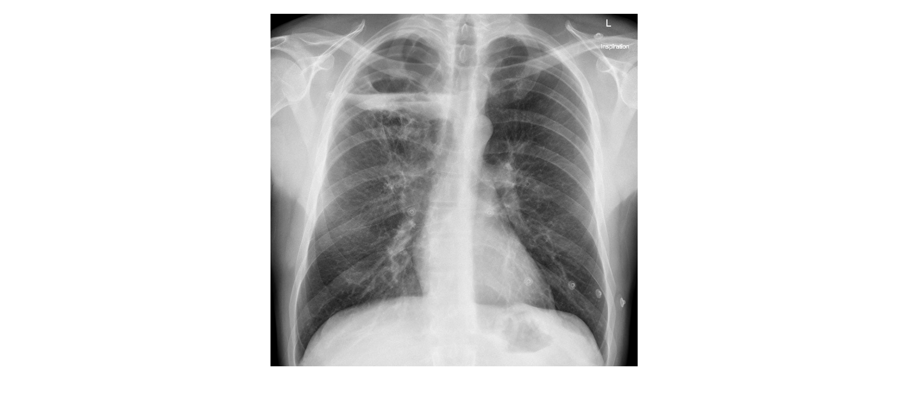

The chest x-ray shows a right apical cystic lucency with fluid level. There is fibrotic streaking extending from the right hilum to the cavity. There is no pneumothorax or pleural effusion.
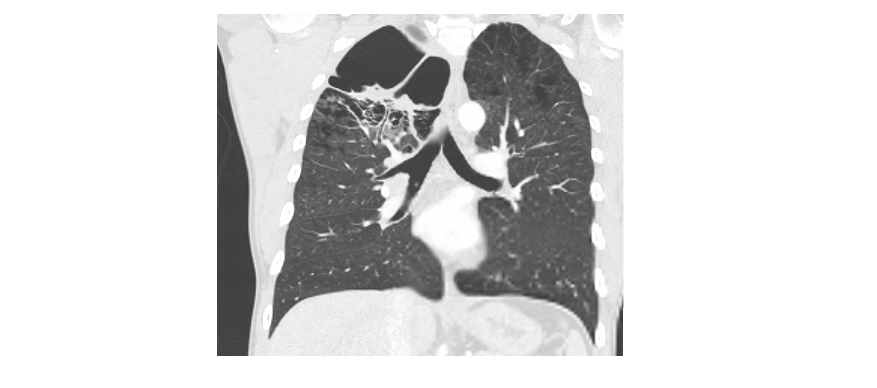
A CT scan showed an infected emphysematous bulla in the right apex with internal septations near the base of the bulla. There were also features of centrilobular emphysema on the CT (high resolution).
 This patient was admitted and treated with intravenous broad spectrum antibiotics and responded favourably.
He had normal alpha one antitrypsin levels and the emphysematous bulla is likely related to the patient’s heavy smoking habit.
Thanks to Dr. Ignatius Munjodzi for the images.
[/peekaboo_content]