
A 55 year old female comes to your ED with a feeling of something stuck in her throat after eating a meal of snapper fish the previous night. She has odynophagia and you perform a lateral neck x-ray looking for a fish bone. The x-ray is here, what can you see?
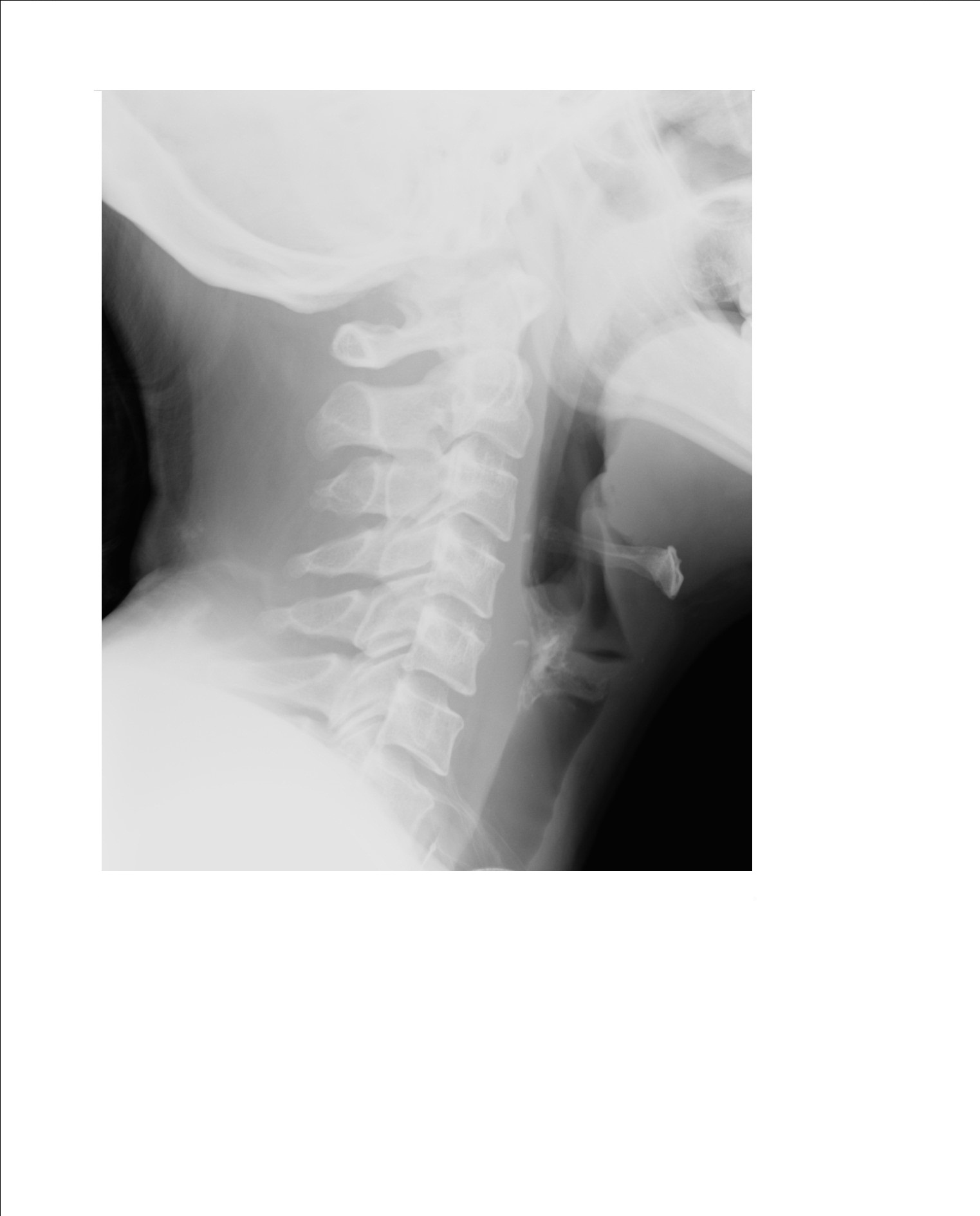
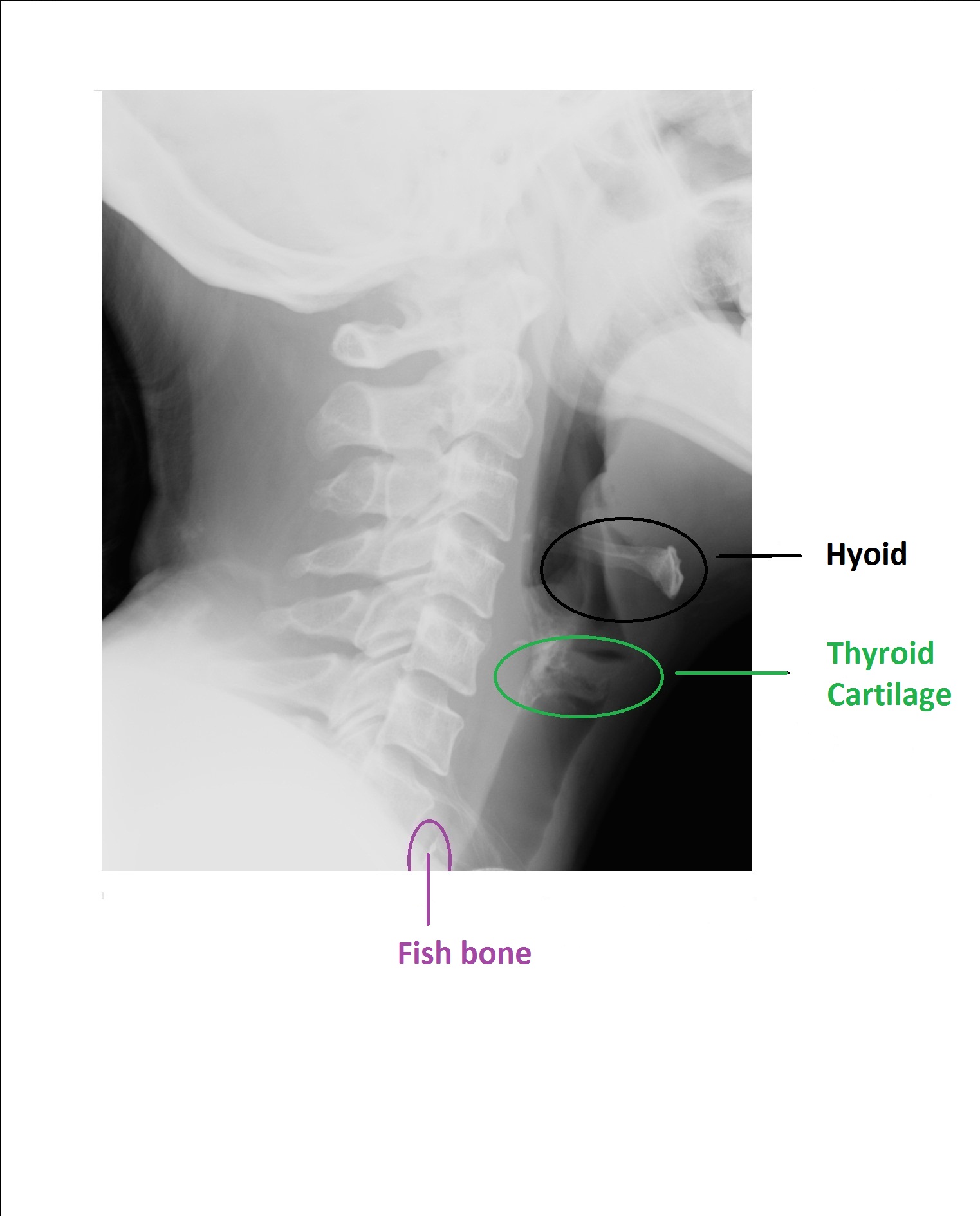
You are slightly confused as there are so many bony bits visible on the x-ray. But if you look carefully, you can spot the linear opaque density in front of the C7 cervical vertebra, right at the bottom of the x-ray surrounded by a pocket of air. There is a fish bone in the cerival oesophagus.
Plain x-ray is not a sensitive test to look for fish bone. Sensitivity is poor, between 30-50% only, and depends on the calcium content in the fishbone. The radio opaque ones are cod, haddock and grey mullet and the least opaque ones are herring, salmon and mackerel. The bottom line is, if the soft tissue neck x-ray does not show a fishbone, it does not rule it out and the patient should be referred to ENT for endoscopic evaluation.
The common sites of lodgement are base of the tongue, the tonsillar fossae (which can be directly visualised in the ED), and in the neck – vallecula, pyriform fossa and the cervical oesophagus.
Undetected fishbone in the pharynx/oesophagus can cause retropharyngeal abscess, oesophagitis,oesophageal perforation and septic mediastinitis, which can be fatal.
[/peekaboo_content]