
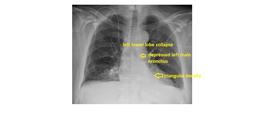
The frontal chest x-ray shows a triangular density behind the heart, silhouetting the medial aspect of the left hemi-diaphragm. The left main bronchus is depressed. These features are suggestive of a left lower lobe collapse.
The lateral view shows increased opacity over the lower thoracic vertebrae (normally should be darker) and posterior displacement of the oblique fissure.
There is also a mild left sided pleural effusion.
This patient had a temperature and elevated inflammatory markers. He was treated with intravenous antibiotics and got better.