
The frontal and lateral thumb x-rays show a lucent area involving the distal phalanx with irregular dorsal cortex. The patient went on to have a CT of the left thumb, which showed lytic destruction of the distal phalanx, with a sequestrum confirming osteomyelitis.
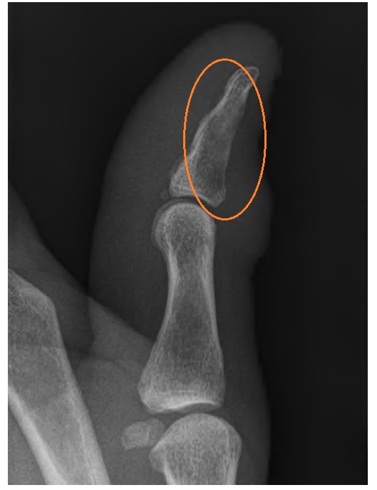
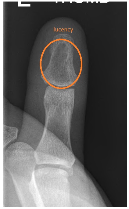

Blood cultures were negative. A bone biopsy revealed staphylococcal growth. The patient was treated with long-term intravenous flucloxacillin as per sensitivities.