
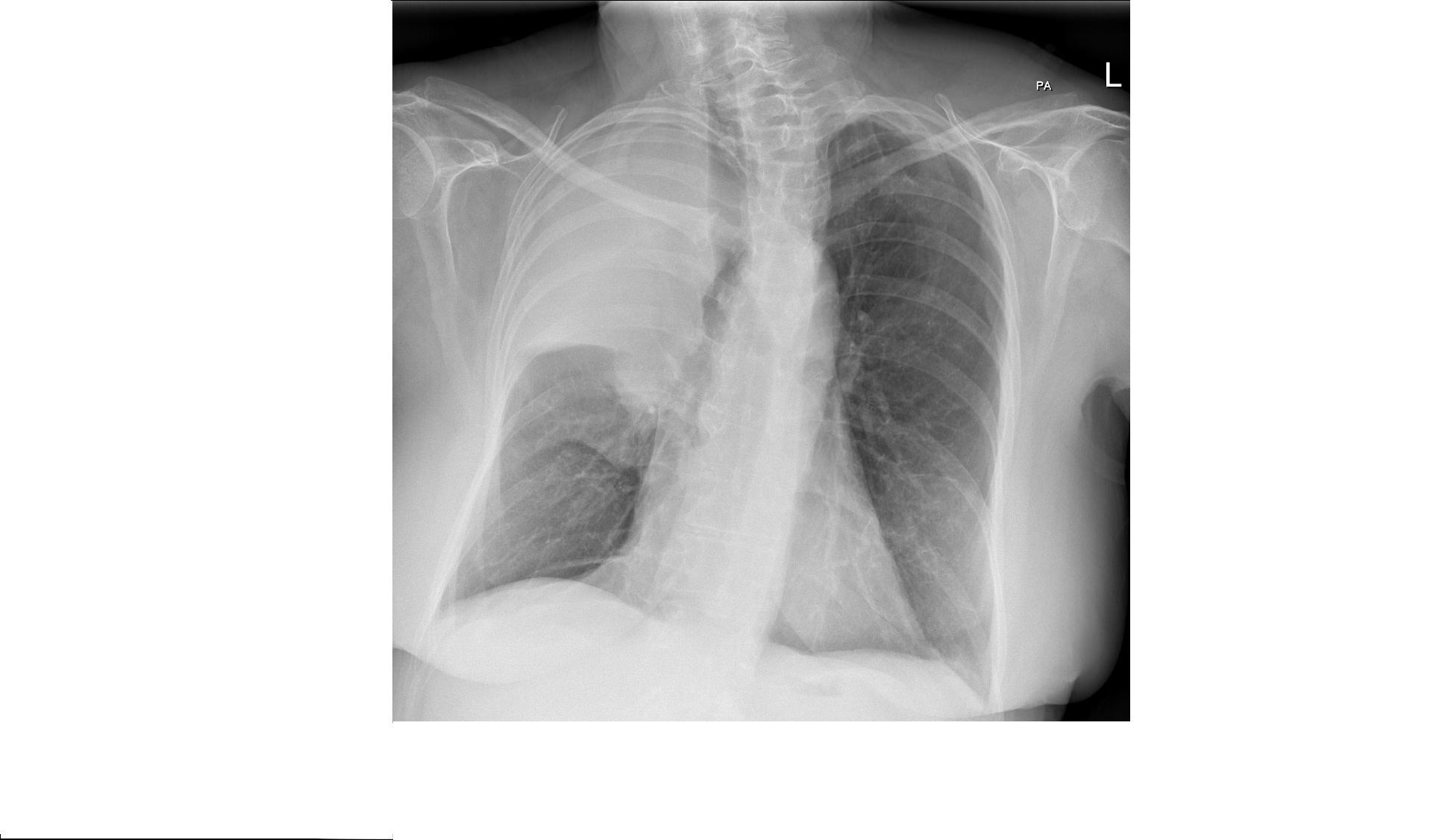
This chest x-ray is from a 60 year old female patient presenting with chief complaints of dyspnoea. She has had her symptoms for few weeks now and denies fever. Her only history of note is 20 pack year smoking history. What can you see?
Click to enlarge
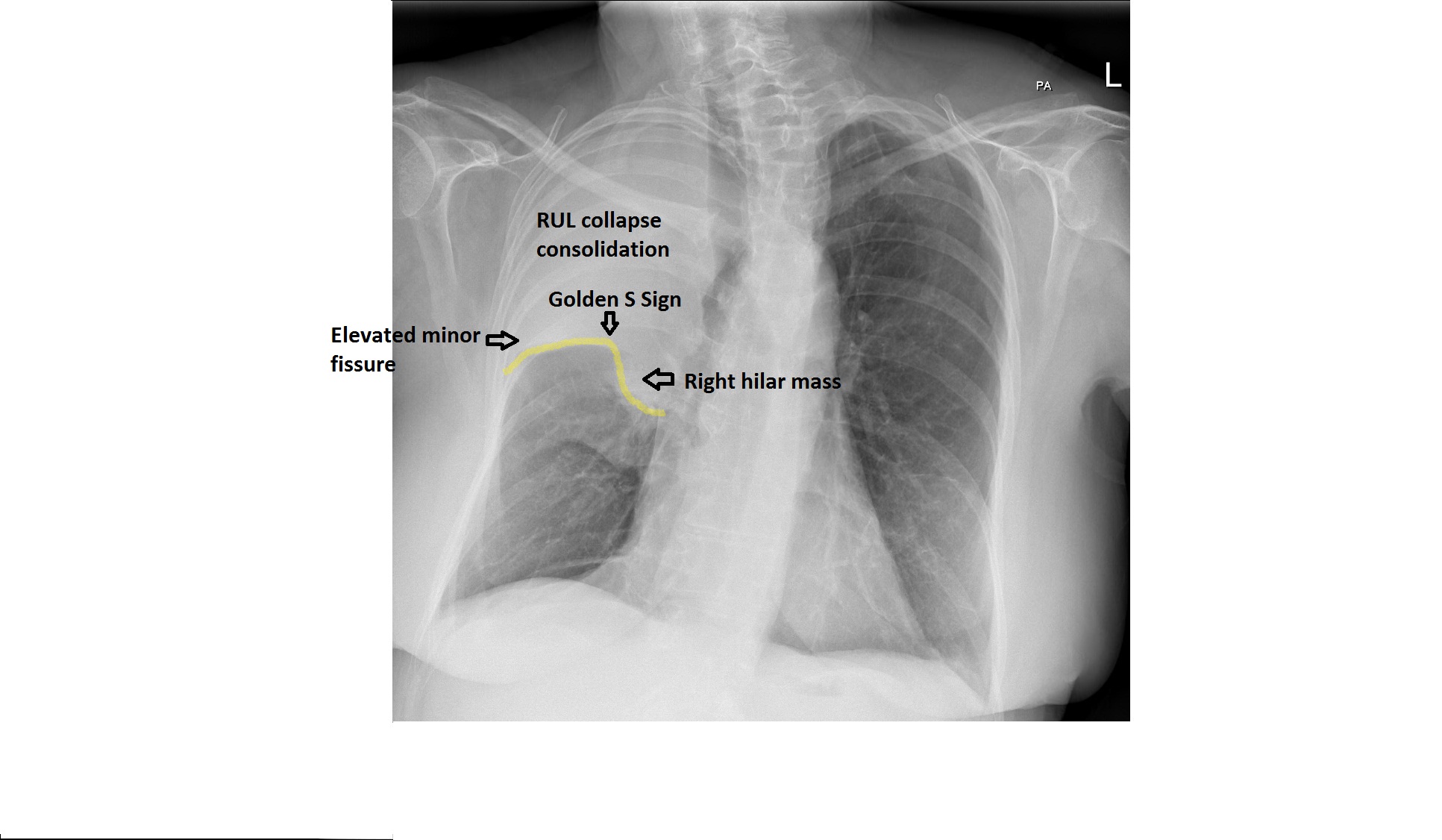
The chest x-ray shows an airspace opacity in the right upper lobe, limited inferiorly by the minor fissure. There is volume loss in the right upper lobe indicated by upward movement of the minor fissure. There appears to be tracheal deviation to the right but the film is rotated. There is no air bronchogram within the opacity. Thre is a right hilar mass likely obstructing the upper lobe bronchus causing collapse consolidation of RUL.
The upward movement of the minor fissure due to collapse and the hilar mass give rise to ‘ Golden S sign’ although this sign is classically described in RUL collapse due to hilar mass.
Click to enlarge