
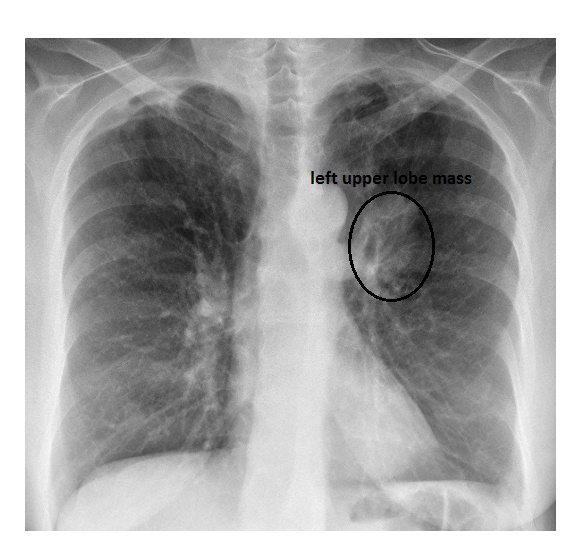
The frontal chest x-ray shows a mass near the left hilum.
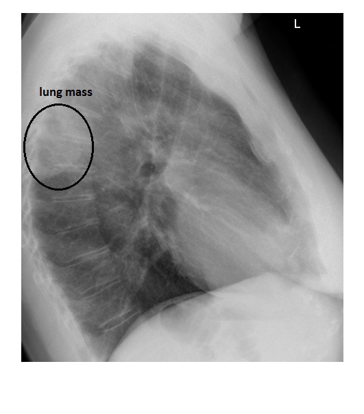
On the lateral chest x-ray, there is an ovoid mass, with irregular margin abutting the pleura in the posterior mediastinum.
The patient had a CT scan of the chest which revealed a primary bronchogenic carcinoma in the apical segment of the left lower lobe. Histopathology demonstrated a small cell carcinoma of the lung.