
The frontal chest x-ray shows retro-cardiac opacity with air bronchogram and silhouetting of the left hemi-diaphragm; in addition, a small left-sided pleural effusion is also present.

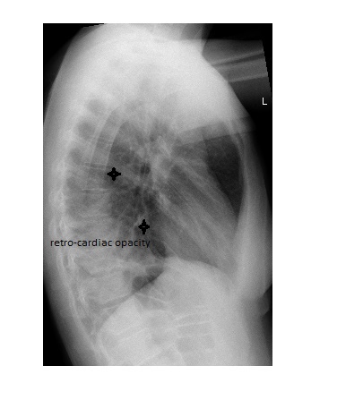
The lateral chest x-ray shows mild displacement of the oblique fissure and posterior cardiac opacity, thus indicating left lower lobe consolidation with partial collapse.
 All patients with pneumonia confirmed on chest x-ray should have a follow-up film in 6 weeks to ensure resolution.