
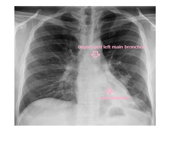
The frontal chest x-ray shows a left lower lobe collapse consolidation. There is retrocardiac opacity, silhouetting of the medial aspect of the left hemidiaphragm indicating left lower lobe pneumonia. There is volume loss in the left thorax as well as depression of the left main bronchus due to left lower lobe collapse.
The patient was treated with intravenous antibiotics, chest physiotherapy and got better.