
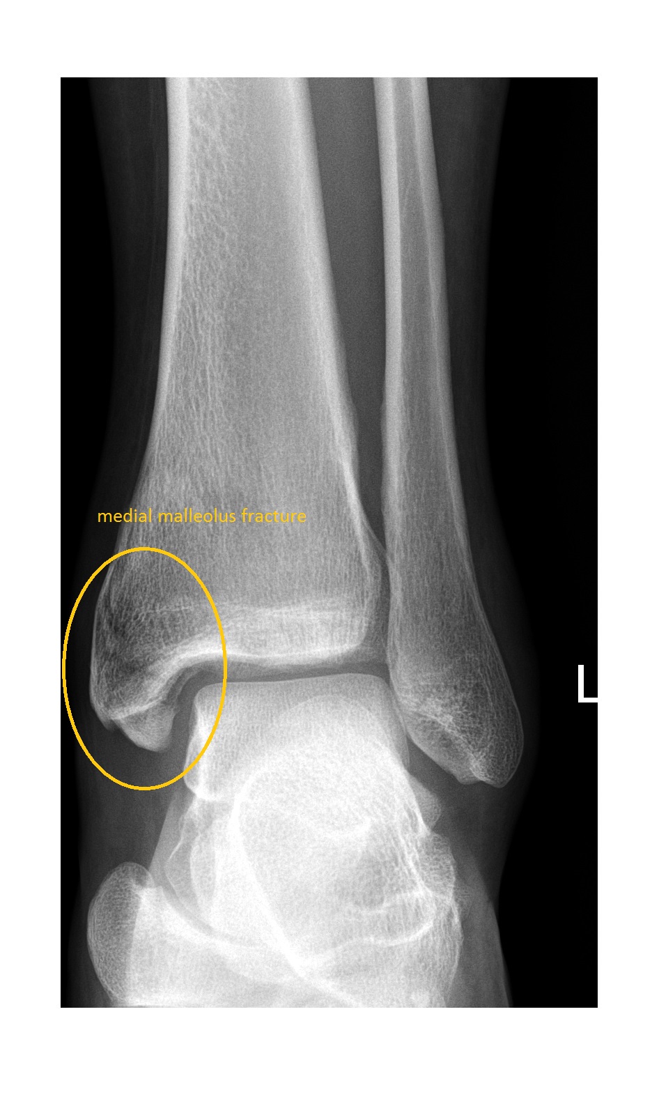
The left ankle AP x-ray shows a comminuted fracture of the medial malleolus. The mortise view of the ankle shows a widened syndesmotic space in addition to the medial malleolus fracture.
The lateral ankle x-ray shows a fracture of the posterior malleolus.


These findings on the x-ray should raise suspicion for a higher fibular fracture and that is what this patient had. This specific injury pattern is called Maisonneuve fracture.

Maisonneuve fracture is an unstable injury and needs referral to orthopaedician for operative management. Always think of Maisonneuve fracture when an ankle x-ray shows isolated medial malleolus fracture or widening of the medial ankle joint space (indicative of a deltoid ligament rupture) and obtain full length leg x-ray to rule out a higher fibular fracture.