
The frontal left wrist x-ray indicates an ulnar styloid fracture. The carpal bones appear crowded with the loss of the uniform 1-2 mm spaces between them.
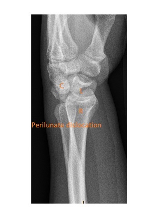
 The lateral view shows a perilunate dislocation. The distal radius and the lunate are in one line with the capitate and the rest of the carpus dislocated posteriorly.
This x-ray is a good example to show satisfaction of search – do not be satisfied with the identification of one injury (ie ulnar styloid fracture).
This patient underwent operative fixation after the initial closed reduction of the dislocation in the ED.
A common mechanism for perilunate dislocation is a fall on an outstretched hand. The lunate remains aligned with the radius but the capitate and rest of the carpus are dislocated dorsally. Perilunate dislocation is commonly associated with other carpal injuries, most commonly a fracture of the scaphoid.
Reference: Grainger & Allison’s Diagnostic Radiology, 7th Edition.