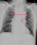
The AP chest x-ray shows a widened mediastinum and depressed left main bronchus, highly suspicious for a traumatic aortic injury. This was confirmed on subsequent CT scan of the chest and the patient underwent a successful repair of their thoracic aorta.
A plain radiograph can have following findings in a patient with thoracic aortic injury:
- Widened mediastinum.
- Obliteration of descending thoracic aorta.
- A left apical cap.
- Downward displacement of the left main bronchus.
- Tracheal deviation to the right.
- Obscurantism of the aortic arch.
- A right paratracheal stripe thickening.
- Deviation of the NG tube to the right.
- Left hemithorax.
- Displaced left or right para spinal line.
- Fractured first rib.
Reference: https://emedicine.medscape.com/article/416939-overview#a2

