
The frontal knee x-ray shows slightly displaced tibial spine fracture. The lateral knee x-ray shows indentation on the lateral femoral condyle (the lateral femoral notch sign). There is an associated knee effusion.
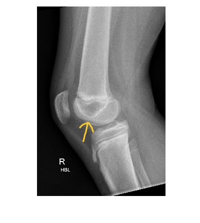
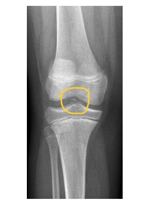
Both injuries are associated with ACL tear and this patient had a 100% ACL tear on a subsequent MRI scan.
The lateral femoral notch sign is characterised by abnormally deep depression of the lateral condylopatellar sulcus. This is due to underlying osteochondral fracture and is highly associated with ACL injury.
Reference: https://pubs.rsna.org/doi/abs/10.1148/radiology.219.3.r01jn12800?journalCode=radiology