
The frontal elbow x-ray shows a lateral epicondyle epiphysis but there is no evidence of medial epicondyle epiphysis in its normal position. There is a bony fragment seen between the olecranon and distal humerus which is likely the displaced medial epicondyle epiphysis.
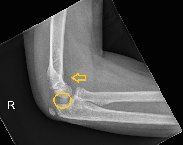
The lateral elbow x- ray shows a large joint effusion. There is an intraarticular bony fragment seen as marked below. This patient has a medial epicondyle avulsion fracture.

The injury was treated with operative fixation.
Medial epicondyle avulsion fractures occur in children as a result of pulling force by the common flexor tendon origin. It can be a part of elbow dislocation.
While interpreting elbow x-rays in children, it pays to remember CRITOE mnemonic which indicates the order of appearance of ossification centres around the elbow joint.
Capitellum – 1 year
Radial head – 3 years
Internal epicondyle – 5 years
Trochlea – 7 years
Olecranon – 9 years
External epicondyle – 11 years
If the lateral epicondyle epiphysis is identified on a paediatric elbow X-ray, one should always be able to visualise the medial epicondyle epiphysis. If not, suspect an avulsion injury to the medial epicondyle epiphysis.
Reference: Grainger & Allison’s Diagnostic Radiology, A Textbook of Medical Imaging, 7th Edition.