
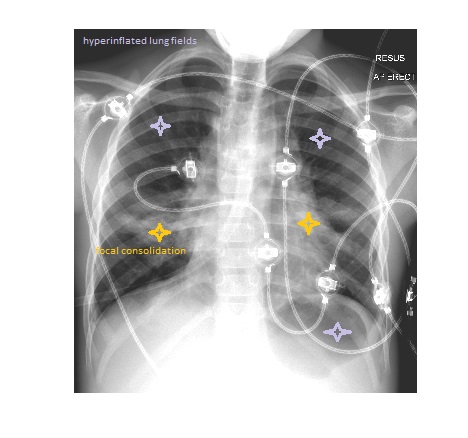
The chest x-ray shows severely hyperinflated lung fields. There is focal consolidation in the right mid and left lower zones. There is gas overlying the left upper quadrant area which should raise suspicion for a pneumothorax but this could also be due to gas trapping in the left lower lung field visible as air underneath the diaphragm.
The child whose presentation was during pre covid era, was managed as critical asthma with
oxygen via face mask
continuous bronchodilators via nebulisation
IV hydrocortisone 4mg/kg
IV aminophylline 10mg/kg over 1 hour
IV magnesium sulphate 0.2 mmol/kg over 20 minutes
IV antibiotics
They got admitted to HDU for further management.
In an asthmatic patient sick enough to be admitted to the hospital, a chest x-ray is required. A chest x-ray may show causes of exacerbation like pneumonia, collapse, or complications of asthma such as a pneumothorax or pneumomediastinum.
Reference: Grainger and Allison’s Diagnostic Radiology, A Textbook of Medical Imaging, 6th Edition.