
The chest x-ray shows a peripheral pleural based irregular pulmonary opacity in the left upper lobe.
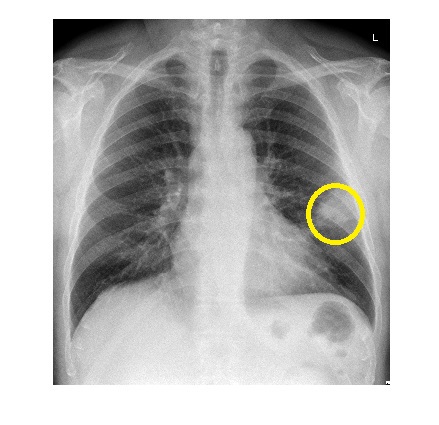
Differentials included –
- Pneumonia
- PE with pulmonary infarct
- Lung malignancy
The patient was febrile with high inflammatory markers. A CTPA ruled out pulmonary embolism. Patient was admitted and treated with intravenous antibiotics.
A chest x-ray done 6 weeks later showed complete resolution of the initial opacity.
A patient with initial diagnosis of pneumonia should have a chest x-ray 6 weeks after antibiotic therapy to ensure resolution of x-ray findings of pneumonia as pulmonary neoplasms like alveolar cell carcinoma can mimic pneumonia initially.