
The chest x-ray shows
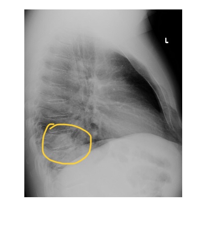
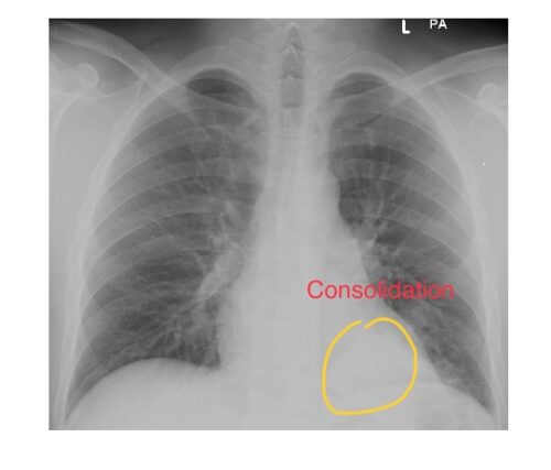
The frontal chest x-ray shows a retrocardiac density/consolidation with air bronchogram. Lateral chest x-ray confirms this area of consolidation in the posterior segment of left lower lobe.
The patient was febrile with respiratory symptoms and had high inflammatory markers and was treated for pneumonia with clinical and radiological improvement. Differential would be lung malignancy (alveolar cell carcinoma).
Repeat chest x-ray in 6 weeks via primary care provider is a must to ensure resolution of consolidation.
Pay particular attention to review areas on a chest x-ray –
- Lung apices.
- Hilum
- Retrocardiac area.
- Below the diaphragm.