
A 30 year old man is brought to your ED by ambulance. He was found lying on his garage floor unconscious by his girlfriend after an earlier argument. He is a NIDDM on Metformin and has some psychological problems on Quetiapine.
Category Archives: Educational Pearls Archive
Ultrasound Post 2 Interpretation.
A registrar happens to do bedside ultrasound on a 55 years old male with abdominal pain and obtains following images.
Answers:
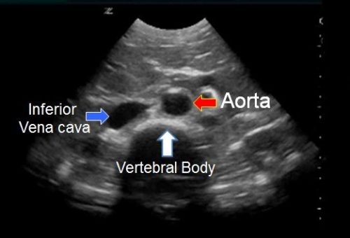
- Above is transvers view of distal aorta , and curvilinear probe has been used. Curvilinear probe is low frequency probe and gives higher depth so ideal for looking at deeper structures hence used in AAA/ AFAST scans.
- Hypoechoic ( black) half circle at the bottom of image is vertebral body shadow which is the main landmark for identifying abdominal aorta. Remember ultrasound waves do not pass through the bones and you get a black shadow as above . Other example is rib shadow when doing FAST scan.
The round structure on right of image ( left of patient ) is Aorta , while the elliptical structure on left of image ( right of patient ) is IVC. Following points will help further to differentiate between two.
a) Aorta lies on left of while IVC lies on right of vertebral body ( patient ).
b) Aorta appears round in shape, while IVC appears elliptical mostly like a tear drop.
c) Both structures can be pulsatile so do not get trapped.
Below is coloured demonstration of above structures.
The daily educational pearl – a bit of history
Any idea when pulse oximetry became available?
The daily educational pearl – Toxic megacolon
Toxic megacolon
= colonic dilation (>6cm) with systemic toxicity, usually due to a acute colitis of different causes. It can involved the whole colon or only one segment.
The daily educational pearl – Darier’s sign
What is Darier’s sign?
The daily education pearl – Argyll Robertson pupils
Argyll Robertson pupils
= bilateral small, irregular, unequal pupils that react to near accommodation but not to light
The daily educational pearl – Injury of the suprascapular nerve
Injury of the suprascapular nerve
This is a rare injury that can occur secondary to:
The daily educational pearl – Injury of the musculocutaneous nerve
Injury of the musculocutaneous nerve
This is a rare injury that can occur secondary to:
The daily educational pearl – recurrent shoulder dislocation
The final post about relocating dislocated shoulders – if you want to teach you patient with recurrent shoulder dislocations how to relocate it themselves:
http://shoulderdislocation.net/relocation/boss-holzach-matter
A patient handout that you can print is available on the site as well.
The daily educational pearl – CUNNINGHAM technique to relocate a dislocated shoulder
I thought we’ll stay with how to relocate dislocated shoulders for now. This is another technique – takes longer, but you don’t need to sedate the patient. It might not work for all patients – especially the ones who can’t control their distress very well.