
A 48 year old female presents complaining of palpitations which started while at gym. The patient has no prior medical history.
Describe and Interpret the ECG
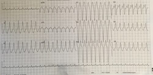
Answer
Rate: 200 bpm
Rhythm: Broad complex regular tachycardia
Axis: Inferior Axis (+90) – use the isoelectric lead method – lead I is the most isoelectric lead
Intervals
PR: no P waves
QRS: 120ms
QTc: 478ms (Fredericia)
Additional:
Typical LBBB pattern, no fusion or capture beats. The precordial transition occurs in V4
The above ECG shows a wide complex regular ventricular tachycardia. Differential for this is a monomorphic ventricular tachycardia, SVT with aberrancy, SVT with aberrant conduction due to WPW, metabolic derangements -hyperkalaemia and sodium channel poisoning.
This ECG has an inferior axis and a typical LBBB pattern and precordial transition >V3, which is consistent with Right Ventricular Outflow Tract Tachycardia. RVOT is usually seen in patients with no underlying structural abnormality and the tachycardia originates in the out flow tract of the right ventricle. Stable patients who present with RVOT can respond to vagal manoeuvres and adenosine.
This patient was treated as a stable monomorphic VT and responded to Amiodarone.
For more reading:
https://litfl.com/right-ventricular-outflow-tract-rvot-tachycardia/
Thank you again to Dr Shenoy for providing the ECG