
The knee x-rays show:
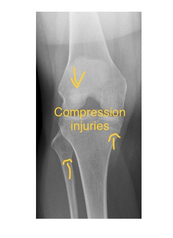
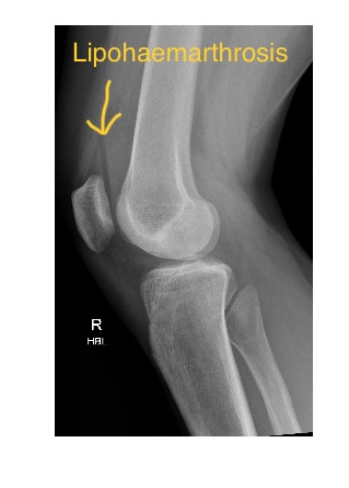
There is a lipohaemarthrosis on the HBL view with AP knee x-ray showing compression type injury in the medial tibial plateau, lateral femoral condyle & neck of fibula.
MRI scan of the knee, in addition to above findings, also showed a complete mid substance ACL tear.
Lipohaemarthrosis in the knee joint is seen as fat fluid level in the suprapatellar bursa on x-ray, best seen on horizontal beam lateral view. This is due to different densities of fat and blood in the joint.
A knee lipohaemarthrosis, when seen, indicates the presence of an intraarticular fracture (tibial plateau, femoral condyle or patellar fracture) and as such, patients with knee lipohaemarthrosis should be referred for specialist management.
Further reading : Grainger & Allison’s Diagnostic Radiology: A Textbook of Medical Imaging, 7th edition.