
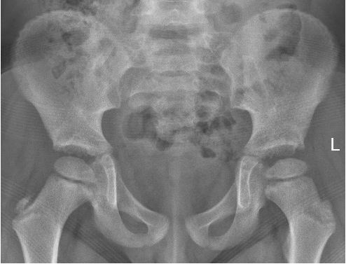
You have just seen a previously well 5 year old with a history of left sided limping. According to the mum, the patient has had this problem for many weeks and the symptoms seem to be getting worse after a trivial fall sustained 24 hours ago. The mother is worried that her son might have sustained a fracture. Pelvic and hip x-rays are as follows; what can be seen? (It is subtle)
Click to enlarge
Click to enlarge

Click to enlarge
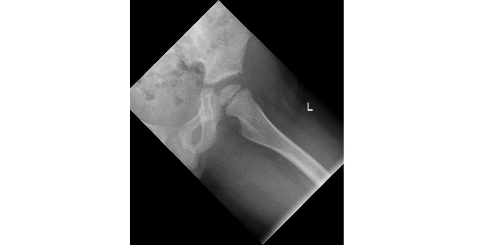
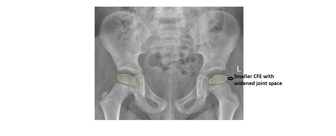
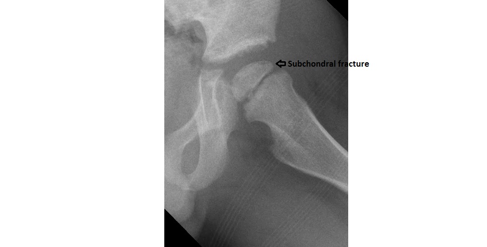
The capital femoral epiphysis is smaller on the left. There is a subchondral linear lucency of the capital femoral epiphysis visible in the frog leg view (represents fracture line). The joint space is slightly widened. Significant sclerosis is not evident yet. The findings suggest avascular necrosis of the femoral head. Given the age of the patient, it is likely due to Perthe’s disease.
When the subchondral lucency is curvilinear in shape, a crescent sign is present.
Click to enlarge
Click to enlarge
Plain x-ray is only about 40% sensitive in detecting early AVN.
Ficat’s staging system of AVN of femoral head based on radiographic findings :-
- Stage 1 – plain x-ray may show minimal osteoporosis and/or poor definition of bony trabeculae.
- Stage 2 – reparative stage showing patchy areas of sclerosis/cysts.
- Stage 3 – crescent sign due to subchondral fracture line; indicates early collapse of the femoral head.
- Stage 4 – flattening of the femoral head and onset of degenerative changes in the joint.
Thanks to Dr. Fiona Beattie for the image.
[/peekaboo_content]