
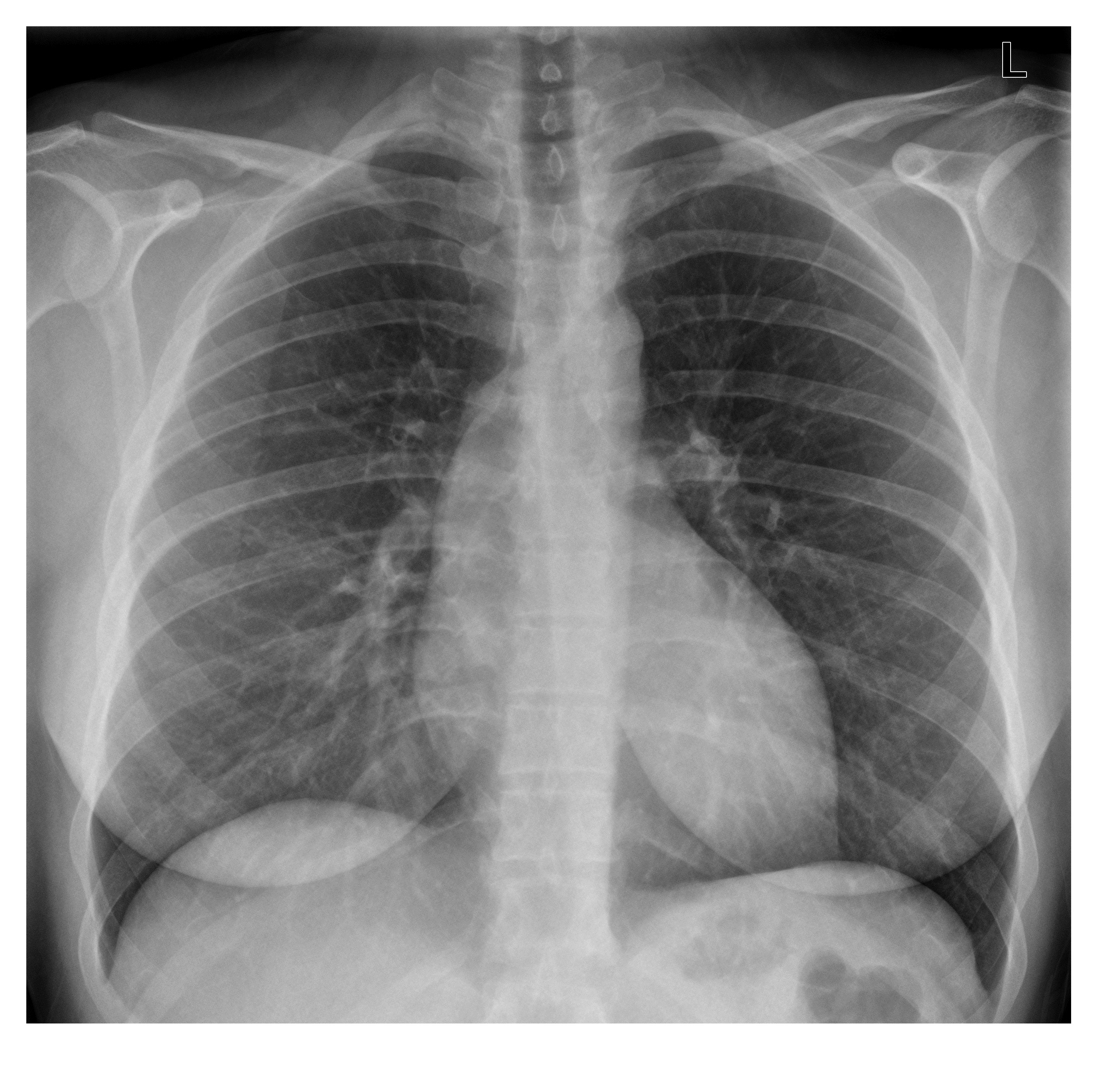
A 34 year old woman who is 3 days post-partum has presented with a sudden onset of central chest pain at 3 am. She has no previous medical illness. As part of investigations, a chest x-ray has been done and is as follows. What is seen on the x-ray? (It is very subtle)
Click to enlarge
At first glance, the chest x-ray appears normal. The lung fields are clear; there is no pneumothorax to account for the sudden onset of pain. However, there is a subtle finding on the cardiac outline which raises the possibility of proximal aortic dissection.
There is a diffuse bulge along the superior aspect of the right heart border, causing focal widening of the mediastinum. This finding is due to aortic root dilatation associated with dissection (Stanford A).

Click to enlarge
This patient had a confirmed proximal aortic dissection on CTA. A slice of axial post contrast CT chest is as follows and shows intimal flap in the mildly dilated ascending aorta (diameter is 4.5 cm).

Click to enlarge
great cxr
I will take a stab at it, since not many ppl being brave on this one 😉
Nothing leaps out other than I think I see a para-trachael line on the right side and possibly opacity at the carina?
Obviously the first thing that leapt into my mind was PE/amniotic embolism and PP cardiomyopathy based on the hx, but I cannot relate those to the findings I think I see – so, keenly waiting on the answer 🙂
Just found answer – wow, nice catch!
What answer did you find Chris? Please let me know 🙂
Hi
Thank you for your comments on the post.
The very reason I chose this case was because it was such an unusual presentation. Chest x-ray findings in this case are subtle, but one should always be on the look-out for minute findings even in a largely normal looking x-ray.
Patients with peripartum cardiomyopathy do not wake up on one fine morning with sudden chest pain; the common pattern of presentation is rather cardiac failure. Yes, they can present suddenly with APO but not with chest-pain.
I do not think there is a right para-tracheal line. If you look closely, the CXR is slightly rotated which may be give off a false impression.
This patient had a undiagnosed bicuspid aortic valve, a risk factor for AD, which unfortunately decided to play up in the post-partum period.