
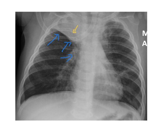
The chest x-ray shows right upper lobe collapse consolidation. There is a triangular density in the right upper lobe, silhouetting the trachea with air bronchogram. The horizontal fissure and the right hilum are pulled up. There is hyperinflation of the lung fields.
The patient tested positive for RSV and underwent conservative treatment for RSV pneumonitis with IV antibiotics and chest physiotherapy.
Lobar collapse with RSV infection is well documented in the literature https://pubmed.ncbi.nlm.nih.gov/4000751/ (I have no subscription to the journal though).
In a child with increased work of breathing from suspected bronchiolitis/viral induced wheeze/asthma, indications for chest x-ray are to look for-
- Pneumonia
- Lobar collapse from mucus plugging
- Foreign body inhalation especially in a toddler
- Pneumothorax
- Pneumomediastinum
- Cardiomegaly/failure (cardiac asthma)