
3 y/o boy admitted via ED for viral induced wheeze as he was requiring ongoing bronchodilator therapy and supplemental oxygen. An ECG was done as child had a rapid pulse.
Q1 Describe and Interpret the above ECG?
Q2 List some electrocardiographic features may be normal in children?
Q3 What are some common indications for performing Paediatric ECGs?
ANS:
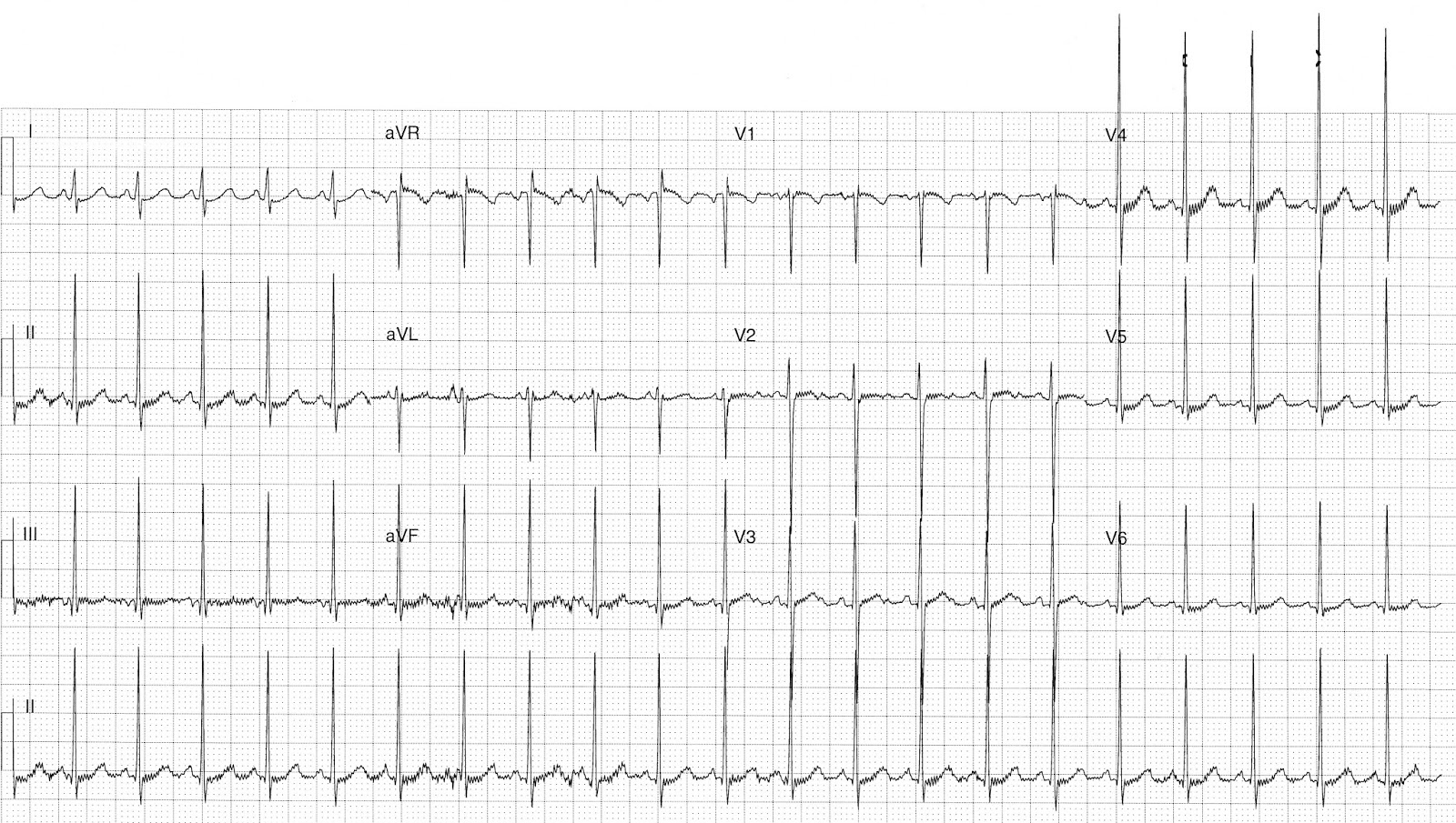
Rate:
- ~126 bpm
Rhythm:
- Sinus rhythm
- Subtle sinus arrhythmia
Axis:
- Normal (~85 deg)
Intervals:
- PR – Normal for age (100ms)
- QRS – Normal (80ms)
- QT – 280ms (QTc Bazette 420 ms)
Additional:
- rSr’ pattern V1
- Baseline artefact
- P wave in II notched but normal height (<3mm) and duration (<90ms)
- U wave in lead V3
- Apparent high voltageds in precordial leads
- Leads II.III,V1,V5,V6 within normal limits for age adjustment
Interpretation:
- Normal age adjusted ECG
ECG features in Children 3-8 years that are normal:
3 – 8 years
- Adult QRS progression in praecordial leads: dominant S in V1, dominant R in V6
- Large praecordial voltages persist
- q waves in left chest leads may be large (<5mm)
- T waves remain negative in right praecordial leads
The following electrocardiographic features may be normal in children:
- Heart rate >100 beats/min
- Rightward QRS axis > +90°
- T wave inversions in V1-3 (“juvenile T-wave pattern”)
- Dominant R wave in V1
- RSR’ pattern in V1
- Marked sinus arrhythmia
- Short PR interval (< 120ms) and QRS duration (<80ms)
- Slightly peaked P waves (< 3mm in height is normal if ≤ 6 months)
- Slightly long QTc (≤ 490ms in infants ≤ 6 months)
- Q waves in the inferior and left precordial leads
Some Common Indications for performing Paediatric ECG in ED:
- Syncope, seizures and “funny turns”
- Cyanotic episodes
- Chest pain or other symptoms related to exertion
- Drug ingestion
- Diagnosis and management of rheumatic fever, Kawasaki’s disease, pericarditis, myocarditis
- Diagnosis and management of arrhythmia
- Diagnosis and management of congenital heart disease
- Family history of sudden death or life threatening event
- Electrolyte abnormalities.
References / Further Reading:
https://pch.health.wa.gov.au/For-health-professionals/Emergency-Department-Guidelines/ECG-interpretation
Thank you Dr Larkin. ECG taken from Dr J Larkin ECG of the Week Blog.