
A 53 year old male presents to ED complaining of chest pain. An ECG is done and is presented to you by one of the nurses:
Describe and Interpret the ECG
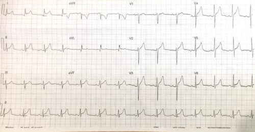
Answer:
Rate: 84 beats/ min
Rhythm: Normal sinus rhythm
Axis: Normal axis
Intervals
- PR 160ms
- QRS 100ms
- QT 376 msec (Bazett)
Additional:
- Global Concave ST elevation, with no reciprocal changes
- ST depression aVR
- STE II>III
- Down sloping TP segment – Spodicks sign
- T waves peaked inferiorly
The above ECG shows typical findings of pericarditis, however in this clinical context (53 year old male) and peaking of T waves I would consider ACS
Further clinical history is required from the patient and serial ECG’s should be done to ensure evolving changes do not occur.
This patient had a previous history of pericarditis 1 year previously, that was treated as a STEMI with a clean PCI, and subsequently diagnosed with pericarditis. His clinical features on this presentation were typical for pericarditis and the patient had serial trops were negative.
For further reading on STEMI vs Pericarditis
https://ecgweekly.com/2016/10/amal-mattus-ecg-case-of-the-week-october-24-2016/
https://litfl.com/spodick-sign/