
You are reviewing 42 year old man who has presented with syncope. His ECG is below.
Interpretation
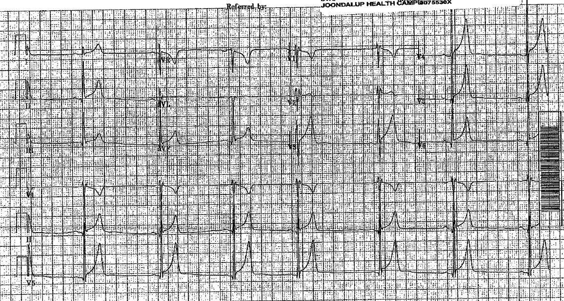
- Rate: 48
- Rhythm: NSR (apologies for the poor quality but seen most easily in lateral leads)
- Axis: Normal (0-90)
- Morphology: large voltage QRS complexes (overlapping in V4-6 – per Sokolov-Lyon criteria S wave V1 and tallest R wave V5-6 >35mm = voltage criteria for LVH, no STD in R sided leads nor R wave peak time>45ms for non-voltage criteria) RSR pattern V1-2, no dagger Q waves in antlateral leads, saddleback STE 1mm seen in V2.
- Intervals: PR 160, QRS 100 R wave peak time <50ms
- Summary: Abnormal ECG in context of syncope (see Ddx below)
Differential Diagnosis:
- LVH
- HOCM
- Possible Brugada Type 3 – this patient went on to have provocation studies to confirm the diagnosis of Brugada.
Further Reading – Textbook:
Chan TC, Brady WJ, Harrigan RA, Ornato JP, Rosen P. ECG in Emergency Medicine and Acute Care. Elsevier Mosby 2005.