
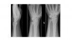
The following x-rays are from a 30 year old male who has presented with left wrist pain after a fall on his outstretched hand. What can you see?
[peekaboo_link name=”Answer”]Answer[/peekaboo_link] [peekaboo_content name=”Answer”]
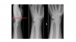
These left wrist x-rays show a fracture fragment on the dorsum of the wrist with adjacent soft tissue swelling, indicating a triquetral fracture. The distal radius, ulna and rest of the carpal bones appear normal.
CT wrist from the same patient :

CT confirmation is not routinely required. In our case, it was done as the patient had mild scaphoid tenderness.
Triquetrum is the second most commonly injured carpal bone after scaphoid, with the relative incidence of a carpal bone fracture being approximately 68% for scaphoid and 18-20% for triquetrum.
The fracture is not evident on the PA view but is visible on the lateral view as an avulsed fragment seen on the dorsum of the wrist.
Treated by below elbow plaster immobilisation and specialist referral.
Reference : Grainger and Allison’s diagnostic radiology
[/peekaboo_content]