
The following OPG x-ray is from a 55 year old patient who has presented to the ED with a high fever and left-sided submandibular pain and swelling. Can you guess the cause? It is very subtle.
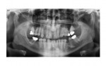
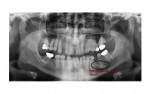
The OPG view shows two ovoid calcific densities near the apex of the lower left 2nd premolar tooth. These represent stones in the left submandibular duct.
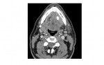
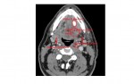
The following axial CT scan slices of the neck at the submandibular level show calcific density at the floor of the mouth on the left side at the expected submandibular duct region. There is soft tissue swelling around the duct in the floor of the mouth, indicating secondary inflammation/infection. The left submandibular gland in this section shows reduced density compared to the right side indicating mild fatty atrophy, likely secondary to the chronic duct obstruction by the calculus.
This patient was admitted under ENT services for intravenous antibiotics. He also underwent transoral removal of the stones.
Thanks to Dr. John Larkin for the images.
[/peekaboo_content]