
The following skull x-ray is from a 60 year old who presented to the ED with vague symptoms of lethargy and weakness. The skull x-ray was performed as part of a skeletal survey following abnormal blood tests. What can be seen?
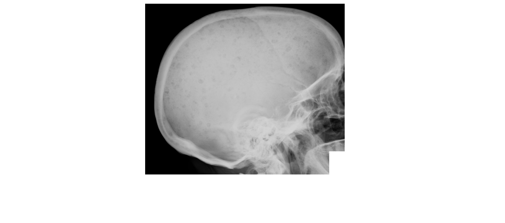
The skull x-ray shows multiple lytic punched-out lesions. These lesions are classic for multiple myeloma and involve the skull and the axial skeleton.
A rare form of sclerotic myeloma is seen in association with POEMS syndrome (Polyneuropathy, Organomegaly, Endocrinopathy, Monoclonal gammopathy and skin changes).
According to Cancer Council Australia, about 1,500 Australians are diagnosed with multiple myeloma each year. The median age of patients is 70 years.
In a patient presenting for the first time to the emergency department, the following abnormalities on blood tests should heighten the suspicion for multiple myeloma. As such, these patients will require prompt admission and a further work up by the haematologist.
- Anaemia – typically normocytic, normochromic; but, can also be macrocytic.
- Renal impairment – due to light chain deposition in the tubules.
- Hypercalcaemia
- Abnormal LFTs – elevated protein and globulin
- Elevated ESR
Reference: http://www.appliedradiology.com/articles/bone-tumors-and-tumor-like-conditions-of-bone & http://www.racgp.org.au/afp/2013/october/multiple-myeloma
[/peekaboo_content]