
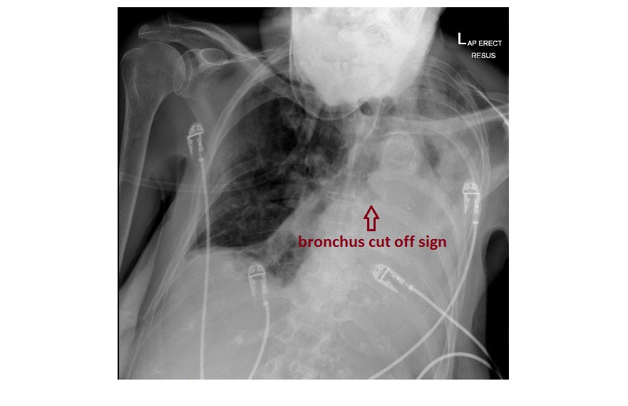
Chest x-ray 1 is an AP film that is technically inadequate. Within the limits, there is a sub-total opacification of the left hemithorax. There is a bronchus cut-off sign on the left with a mediastinal shift to the left. This patient went on to have a CT of the chest which showed a mucus plug occluding the left main bronchus, resulting in near total collapse of the left lung.
 Chest x-ray 2 is from a patient who has previously had a left pneumonectomy. There is mediastinal shift to the left and there are surgical clips visible around the left main bronchus.

Chest x-ray 3 is from a patient with massive left-sided pleural effusion. There is associated mild mediastinal shift to the right side.Pleural plaques are also visible in the right hemithorax. Based on case, cause and complications analysis, this is a case of severe left pleural effusion in a patient with asbestosis, which is likely complicated by mesothelioma.

Causes of white out hemithorax:
- With mediastinal shift towards the affected side – lung collapse, previous pneumonectomy
- With no mediastinal shift – consolidation (will be associated with air bronchogram sign)
- With mediastinal shift to the opposite side – massive pleural effusion