
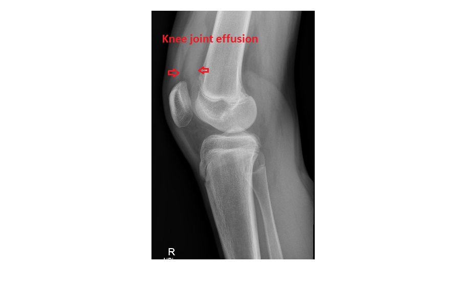
The frontal knee x-ray shows an elliptical fragment of bone lying next to the lateral tibial plateau; this is known as a Segond fracture. The HBL view shows knee joint effusion.

Segond fracture may be a small avulsion fracture but it signifies a significant internal derangement of the knee joint. In adult patients, it is associated with an ACL tear in 75-100% of cases. It is also linked to meniscal injury. A common mechanism of the fracture is internal rotation and varus stress to the knee joint in flexion.
In adults, a Segond fracture occurs in approximately 9-12% of all ACL tears. In skeletally immature patients, the link between ACL injury and Segond is not as definitive due to differences in the strength of bone and ligaments (i.e. compared to bone, ligaments are stronger in children). But none the less, all cases require a referral to an Orthopaedician for further management.
This particular patient underwent a MRI scan of the knee and was noted to have an avulsion of the tibial attachment of the ACL (Segond fracture), a posterolateral capsular injury and bony contusions involving the lateral tibial and femoral condyles.
Reference article can be found here