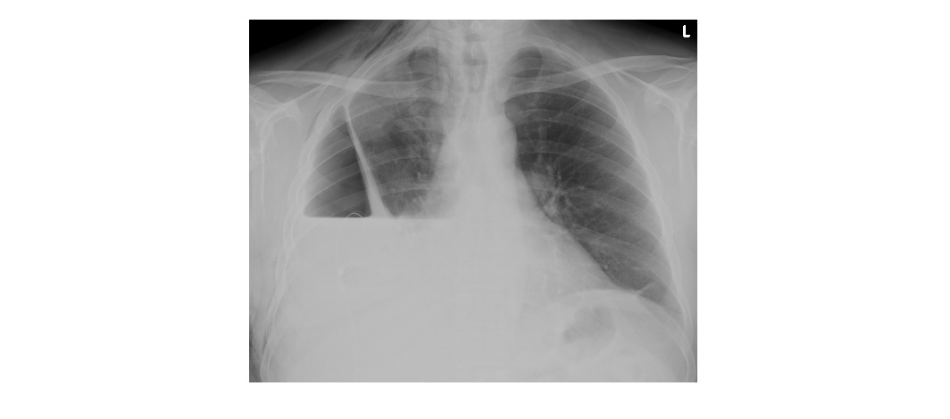
The following chest x-ray is from a 40 year old with right sided chest pain and dyspnoea. This x-ray has been taken the day after patient has had a chest drain insertion. What can be seen?
Answer will be posted on 18/11/2015.

The following chest x-ray is from a 40 year old with right sided chest pain and dyspnoea. This x-ray has been taken the day after patient has had a chest drain insertion. What can be seen?
Answer will be posted on 18/11/2015.
R hydropneumothorax. chest drain in an unusual position, does not appear to have been inserted laterally. curled back on itself at the end but not in a way that suggests it is a pigtail catheter. no definite kinking identified.
ddx: blocked ICC (eg inspissated secretions or ICC too narrow if exudate or blood is being drained)
kinked ICC (internally or externally)
underwater seal drain not attached properly
ICC in s/c tissue not pleural space – (r/v films from time of insertion & technique of insertion).
If ICC cannot easily be unkinked or unblocked, needs new ICC left midaxillary line 5th intercostal space confirming finger through to pleura.