The chest x-ray shows:
- A focal protrusion, not silhouetting the left heart border, projected over the proximal descending thoracic aorta. On the lateral chest x-ray, this bulge is appreciated as an increasing density over the mid-thoracic spine, adjacent to the descending aorta. The location of the protrusion raises the possibility of a focal aneurysm of the thoracic aorta.
- A solitary pulmonary nodule measuring approximately 15 mm, seen in the right lower zone.

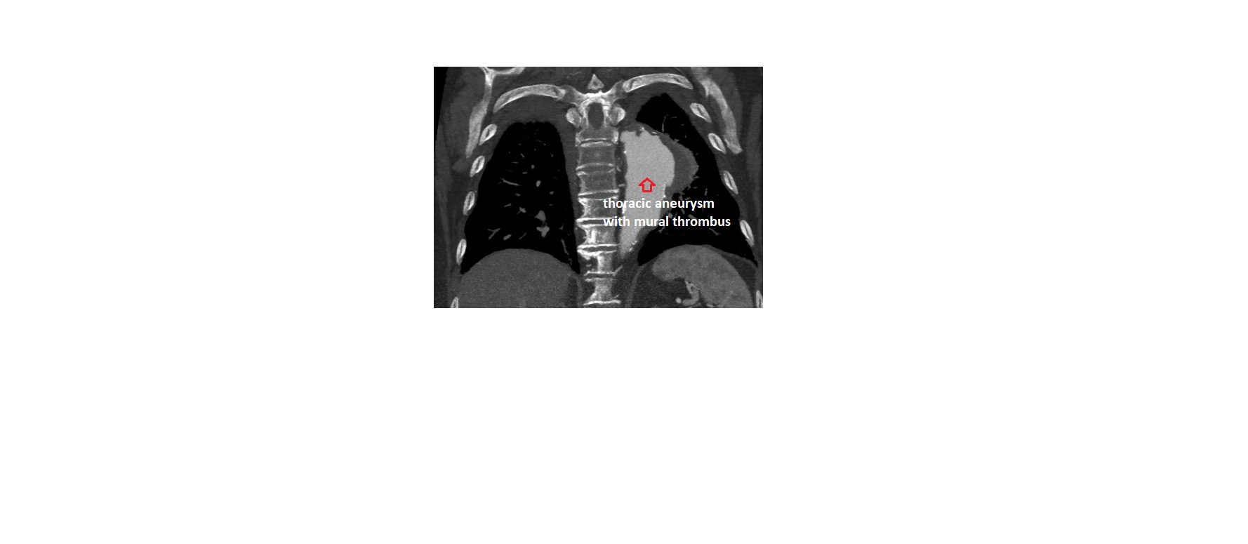
This patient went on to have a CT aortogram, which confirmed a descending thoracic aortic aneurysm, measuring 7cm, with mural thrombus. On the CT, the solitary pulmonary nodule did not show features of malignancy.