
Your next patient is a 68 year old female with mild dyspnoea. On examination, she is stable with mild epigastric tenderness only. You obtain chest x-ray as part of your investigation and the x-rays are here.

Click to enlarge
Looking at the PA view, you are beginning to wonder if you are dealing with a left diaphragmatic hernia. Here is the lateral view. What can you see?
[peekaboo_link name=”Answer”]Answer[/peekaboo_link] [peekaboo_content name=”Answer”]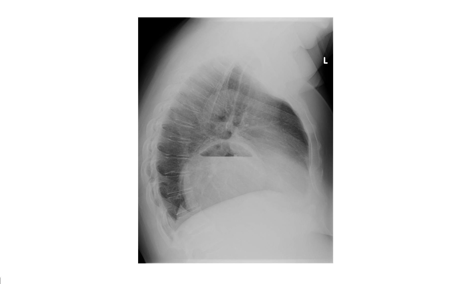
Click to enlarge
The PA chest view shows distended stomach with air fluid level in the left hemithorax. The lateral view shows fluid/air filled stomach in retrocardiac position. There is a large hiatus hernia.
Hiatus hernia can be of 2 types :
- Sliding – Commonest, the gastro-oesophageal junction is above the diaphragm. Predisposes to reflux, oesophagitis.
- Para oesophageal – Portion of the stomach lies above the diaphragm after herniating through the oesophageal hiatus but the gastro-oesophageal junction lies below the diaphragm. Can lead to life threatening complications like volvulus or strangulation.