
It is 2 am and you have just seen a 30 year old female patient who just twisted her ankle at a party and is unable to weight-bear. Her ankle is swollen and painful and you decide to do an x-ray of her ankle and the films are as follows. What can you see?
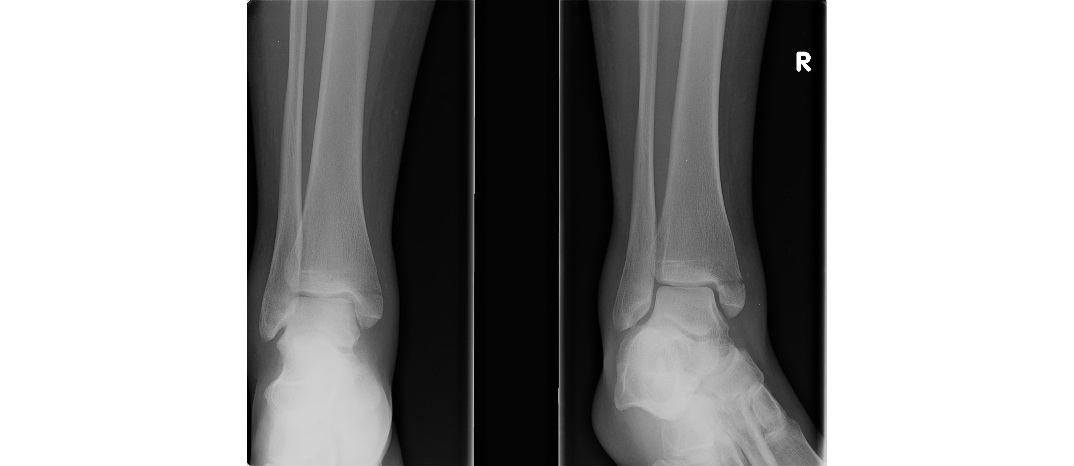
You are happy that her x-ray correlates with your clinical diagnosis of medial malleolar fracture. There is an undisplaced transverse fracture of the medial malleolus. While reassessing the patient, you notice that the patient has got tenderness over the upper leg as well. So, you obtain additional x-rays of her knee. What can you see?
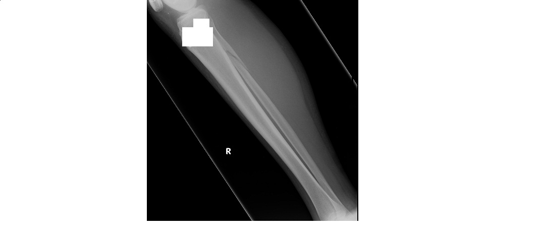
There is a spiral, minimally displaced fracture of the upper third of the fibula. In other words, this patient has Maisonneuve’s fracture.
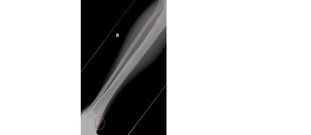
Maisonneuve’s fracture is a combination of proximal fibular fracture and medial malleolar fracture or disruption of the deltoid ligament. The injury force is transmitted through the interosseous membrane to the level of fibular fracture. It usually results from eversion/external rotaion injury to the ankle. It is an unstable injury, hence the right diagnosis is very important.
Suspect Maisonneuve’s fracture when an ankle x-ray shows:
- Medial malleolar fracture
- Widening of the distal tibio-fibular syndesmosis (the tibio-fibular clear space, when measured 1cm above the tibial articular surface, should be less than 5mm in width)
- Lateral talar displacement
Treatment is mostly surgical with operative fixation of the medial malleolus with syndesmotic screws; hence patient should be referred to ortho but referral can wait till 7am. But treat with back slab, leg elevation, analgesia etc in the meanwhile.
The bottomline is not to forget examining the entire leg in patients with twisting injury to the ankle as ankle pain can be the only presenting complaint.
[/peekaboo_content]