
The frontal elbow x-ray shows a lateral condylar fracture of the humerus with approximately 3 mm separation of the fracture fragments.
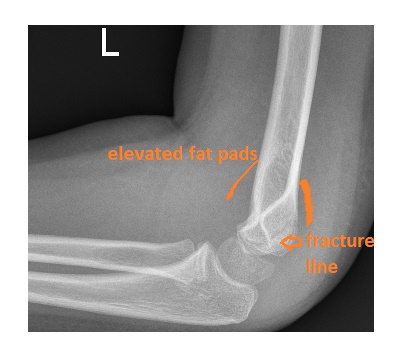
The lateral view shows a large effusion in the elbow joint with an elevated anterior and posterior fat pad. The fracture line through the distal humerus is visible.

Lateral condylar humeral fracture is the 2nd common elbow injury after the supracondylar fracture. It is important to recognise this fracture in the ED and refer patients to the orthopaedic team as complication rate is high (delayed union, non-union and growth disturbance)
Further information including classification and treatment can be found here.