
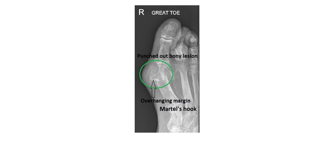
The left foot x-ray shows erosion at the medial aspect of the great toe proximal phalanx base. The margins are overhanging and joint space is narrowed. There is no peri-articular osteopenia or sclerosis.
In the context of diabetes and cellulitis, septic arthritis of the metatarsophalangeal joint (MTP) of the great toe is a likely possibility. Gout involving the MTP joint of the great toe may also have a similar appearance; here is an x-ray with a confirmed gouty arthritis of the MTP joint of the great toe (Imaging Case of the Week 175).

The patient had a bone scan as well as MRI of the left foot which revealed septic arthritis of the MTP joint of the great toe and osteomyelitis of the head of the 1st metatarsal.