
It is 3 am and you are about to see a 38 year old female patient brought in by paramedics with complaints of dyspnoea. Her vital signs are bit concerning; she is tachypnoeic with a respiratory rate of 30 and her O2 sats are 90 % in room air. As you begin talking to her, she says she wants to go home. You convince her to get a chest x-ray and she reluctantly agrees. Here is the PA chest x-ray. What can you see ?
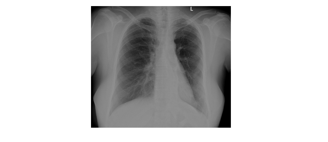
Click to enlarge
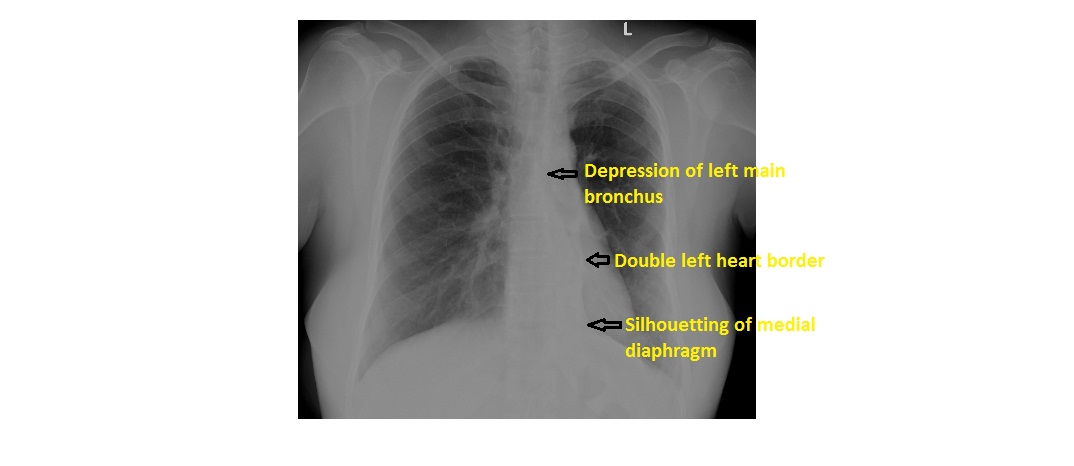
The chest x-ray shows left lower lobe collapse which gives rise to the double left heart border or the sail sign.
- There is a triangular density behind the heart, silhouetting the medial aspect of left diaphragm.
- The second left heart border is due to the edge of the collapsed lower lobe.
- There is volume loss in the left hemithorax with elevation of the left hemidiaphragm and depression of the left main bronchus.
Click to enlarge
Left lower lobe collapse can be easily missed if careful attention is not paid at the time of interpretation, especially in an underpenetrated film. Unfortunately, I do not have a lateral view for this patient.
[/peekaboo_content]