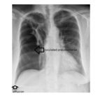
The frontal chest x-ray shows a large area of lucency involving the right lung. There is an associated small right pleural effusion and atelectasis involving the right upper lobe.
Other findings include horizontally placed ribs and a hyper-expanded left lung field.
Based on the findings, the differentials could be a large emphysematous bulla involving the right lung or a right-sided pneumothorax. The patient went on to have a CT scan of the chest, which indicated a loculated right pneumothorax.
The patient was managed conservatively without a chest drain and underwent serial x-rays. A follow up chest x-ray at 5 weeks showed complete resolution of the pneumothorax.
