
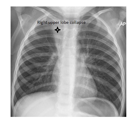
The chest x-ray shows peribronchial thickening. There is an area of triangular density in the right upper zone with the apex towards the right hilum. The right hilum has been pulled up. The overall appearance is suggestive of bronchiolitis, complicated by a right upper lobe collapse. The oxygen tubing is visible as an external artifact.
The patient was managed conservatively.