
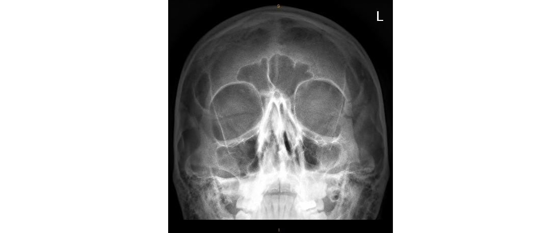
It is 1 am on a Saturday night. You are about to see a 30 year old man who has been assaulted at a pub. The left side of the patient’s face is swollen and the facial x-rays are as follows. What can you see?
Click to enlarge
Click to enlarge
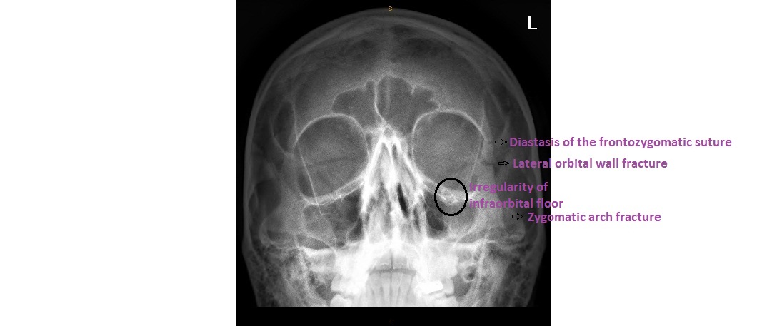
The OM (occipitomental) view shows:
- irregularity over the centre of the left orbital floor
- slight widening of the left frontozygomatic suture
- fracture of lateral orbital wall
- maybe fluid (blood in the context of trauma) in the left maxillary antrum
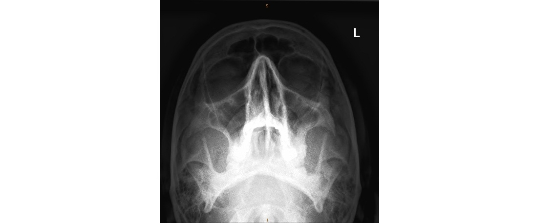
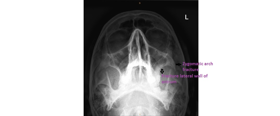
The OM (30) view shows:
- left zygomatic arch fracture
- there is also step deformity (inwards) of the lateral wall of the maxillary antrum
This patient has a combination of injuries called the Tripod Fracture which essentially consists of ipsilateral triad of:
- zygoma fracture (break through the orbital floor and lateral wall of antrum)
- zygomatic arch fracture
- widening of frontozygomatic suture
Click to enlarge
Click to enlarge
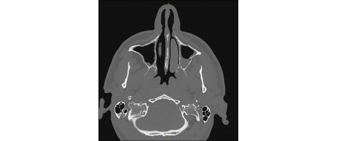
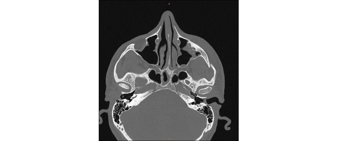
Here are the CT images showing the left infraorbital fracture and fluid in the left maxiallry antrum clearly:
Click to enlarge
Click to enlarge
This patient needs referral to maxillofacial surgeons from the ED.
Thanks to Dr. Michael Lovegrove for the images.
[/peekaboo_content]